Advanced Imaging Services
Comprehensive diagnostic technology for precise orthopedic care at Pacific Crest Orthopedics
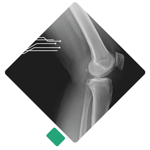
Fast, Low‑Radiation X‑Rays
We believe that the sooner you have answers, the sooner you can heal. Our practice maintains both digital X‑ray and mini‑fluoroscopy machines on‑site, which means we can capture high‑quality images during your appointment without sending you elsewhere. These machines are designed to deliver low‑dose radiation while providing clear, detailed images of bones and joints.
Digital X‑rays are commonly used to evaluate:
-
Clavicle, shoulder, and elbow injuries
-
Wrist, hand, and finger fractures
-
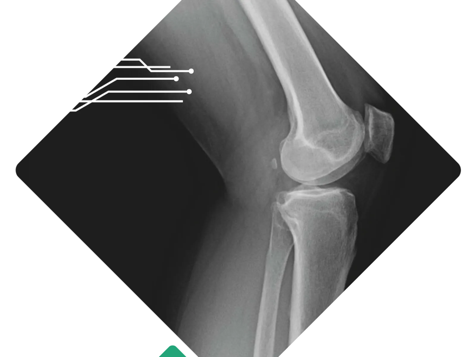
Hip, femur, knee, and leg trauma
-
Tibia/fibula, ankle, and foot injuries
At our practice, your images are reviewed right away so you can talk through the findings and next steps before you head home. In orthopedics across the United States, requesting an X‑ray before your visit is considered standard care. It allows our specialists to see your bones and joints clearly and makes the best use of your time when you come in. We understand some people feel uneasy about imaging, but it's a routine part of musculoskeletal care that's focused on getting you the most accurate evaluation. If you are pregnant or scheduling an appointment for a child, please let us know; we'll coordinate with your doctor and take any extra precautions needed to keep you safe.
At Pacific Crest Orthopedics we have both X-ray and fluoroscopy machines in the office for quick diagnosis and evaluation of all orthopedic injuries. The machines are low radiation and give high-quality images. We can take immediate X-rays of most orthopedic injuries including injuries to the clavicle, shoulder, elbow, wrist, hand, finger, hip, femur, knee, tibia/fibula, ankle, and foot. Each of these can be taken during your appointment and then read promptly by the provider for an efficient diagnosis.
At Pacific Crest Orthopedics, we have advanced X-ray machines right here in our office that use very low radiation to produce clear, detailed images of your bones and joints. We can take X-rays of many areas, including your collarbone, shoulder, elbow, wrist, hand, fingers, hip, thigh, knee, shin, ankle, and foot, all during your appointment. This allows your doctor to quickly review the images and provide an accurate diagnosis so treatment can begin promptly.
As an orthopedic practice specializing in muscles and bones, requesting an X-ray before your visit is a standard part of our care to ensure the best possible evaluation. While some patients may feel hesitant about this, please don't be alarmed; this is routine and designed to help us serve you better. For pregnant and pediatric patients, we take extra precautions and will consult with your provider before proceeding with any X-rays.
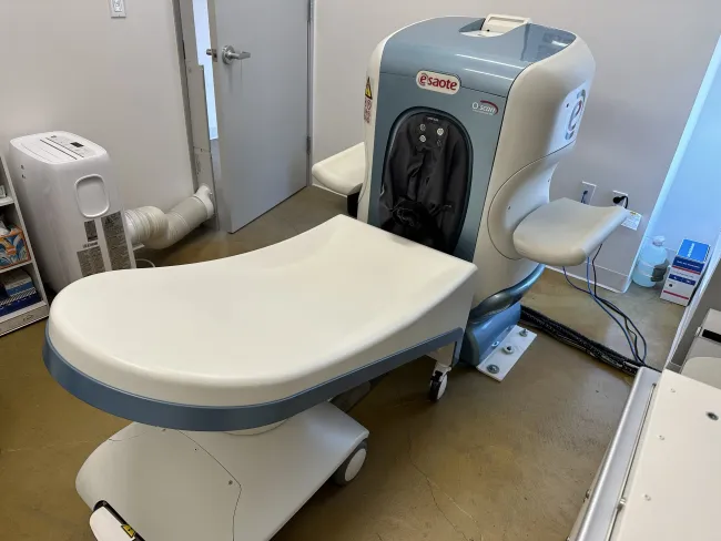
MRI Services: Advanced Soft Tissue Imaging
When X-rays provide initial insights but your provider needs a deeper understanding of soft tissue structures, magnetic resonance imaging (MRI) becomes an essential diagnostic tool. MRI technology offers unparalleled visualization of ligaments, cartilage, menisci, tendons, and other soft tissue structures that standard X-rays simply cannot reveal.
On-Site Convenience
Our Mission location features an advanced MRI machine suitable for imaging the elbow, wrist, knee, ankle, and foot. This means fewer trips to external facilities and more streamlined care coordination.
Non-Invasive Technology
Unlike CT scans or X-rays, MRIs use radio waves and magnetic fields instead of ionizing radiation, making them a safer option for detailed soft tissue evaluation. The procedure is completely non-invasive and painless.
Insurance Navigation
Most insurance plans require X-ray evidence before approving MRI authorization. Our team handles the prior authorization process, which typically takes 10-12 business days depending on your individual insurance plan.
Flexible Payment Options
We offer competitive cash pay rates for MRI services if you prefer to bypass insurance authorization or don't have coverage. Contact our office for current pricing and availability.
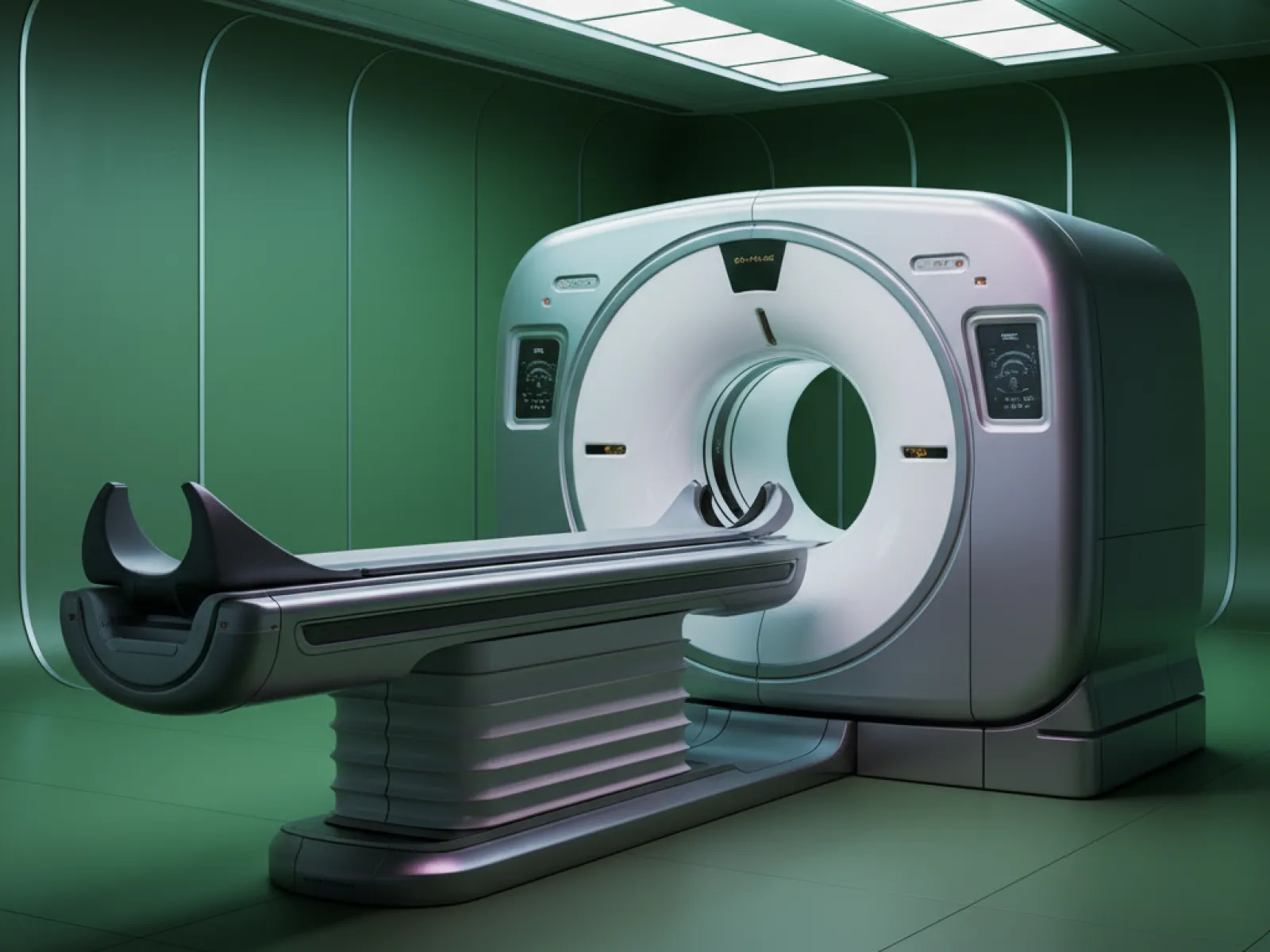
CT Scans: Detailed Fracture Analysis
When Precision Matters Most
Computed tomography (CT) scans provide three-dimensional, highly detailed images that help your provider understand complex fractures with exceptional clarity. Think of CT scans as significantly more detailed X-rays that reveal bone structures from multiple angles and perspectives.
While CT technology does use ionizing radiation similar to X-rays, the level of detail it provides is often essential for making critical treatment decisions, especially when surgical planning or precise measurements are required.
Ultrasound-Guided Injections
Ultrasound technology at Pacific Crest Orthopedics extends beyond diagnostic imaging. Our providers utilize ultrasound guidance for therapeutic injections, combining real-time visualization with precise treatment delivery for optimal results and patient safety.
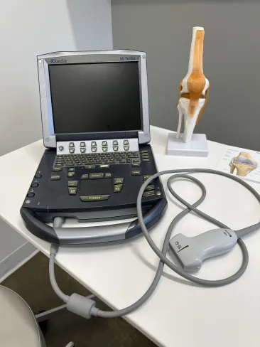

Enhanced Accuracy
Real-time ultrasound imaging allows your provider to visualize muscles, tendons, ligaments, and joints during the injection procedure, ensuring medication reaches the exact target location for maximum therapeutic benefit.

Reduced Risk
By providing continuous visualization of anatomical structures and the needle path, ultrasound guidance significantly reduces the risk of complications and ensures the injection avoids sensitive structures like nerves and blood vessels.

Minimally Invasive
Ultrasound imaging is completely non-invasive and uses harmless sound waves rather than radiation. The procedure is performed in-office with minimal discomfort and no recovery time required.
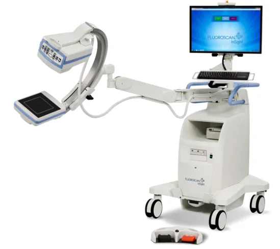
Fluoroscopy: Real-Time Motion Imaging
In addition to standard X‑rays, we utilize a fluoroscopy machine in select office locations. Fluoroscopy uses a continuous low‑dose X‑ray beam to produce live video images of bones, joints, and soft tissues. This allows your provider to watch structures move in real time, making fluoroscopy valuable for: